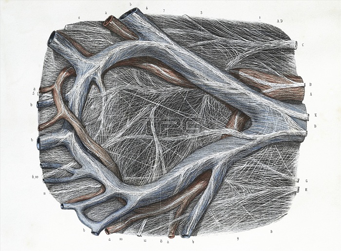
Mesocolon. 19th century artwork showing the veins (blue), arteries (red) and nerves (white) of the mesocolon. The mesocolon is part of the peritoneum, the layer of connective tissue that lines the abdominal cavity. The mesocolon functions to attach the colon (large intestine) to the posterior abdominal wall. This anatomical artwork is Fig 1 of plate 51 from volume 5 (1839) of 'Traite complet de l'anatomie de l'homme' (1831-1854). This 8-volume anatomy atlas was produced by the French physician and anatomist Jean-Baptiste Marc Bourgery (1797-1849). The illustrations were by Nicolas-Henri Jacob (1781-1871).
| px | px | dpi | = | cm | x | cm | = | MB |
Details
Creative#:
TOP14051391
Source:
達志影像
Authorization Type:
RM
Release Information:
須由TPG 完整授權
Model Release:
N/A
Property Release:
No
Right to Privacy:
No
Same folder images:

 Loading