
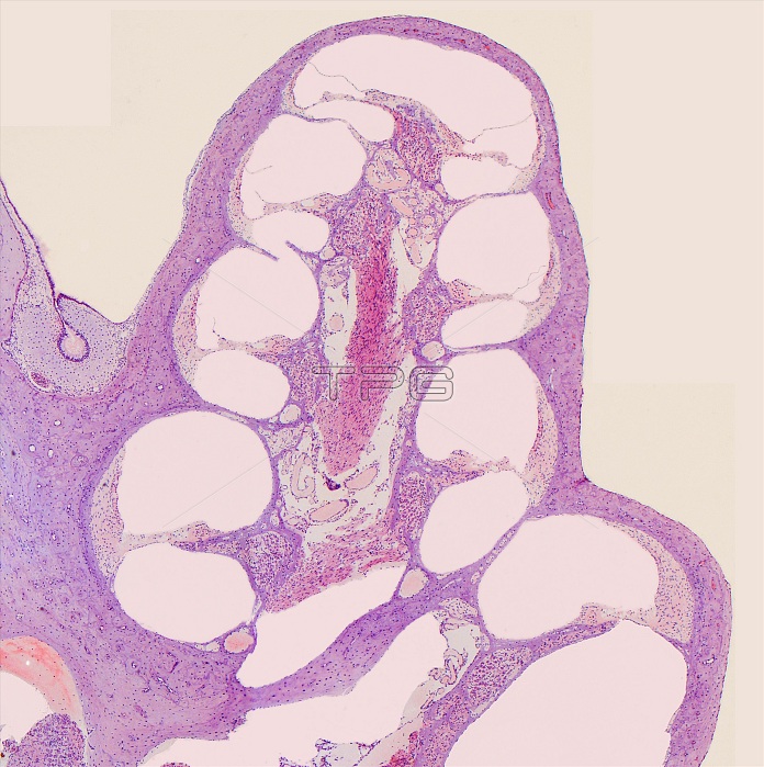
Light micrograph (LM) of a section through the cochlea. The cochlea is a tapering, fluid-filled spiral cavity 3.5cm long. Two membranes run the length of the spiral & divide it into 3 parallel ducts. In each coil, the upper membrane is tissue- thin; the lower, or basilar membrane, is thicker. The ducts they form are easily seen. In the middle duct, on the basilar membrane, is the Organ of Corti, where sensory hair cells react to sound & convert it into electrical signals which travel to the brain via the cochlear nerve. Magnification: x100MB at 10cm size.
| px | px | dpi | = | cm | x | cm | = | MB |
Details
Creative#:
TOP13822187
Source:
達志影像
Authorization Type:
RM
Release Information:
須由TPG 完整授權
Model Release:
N/A
Property Release:
No
Right to Privacy:
No
Same folder images:
 Loading