
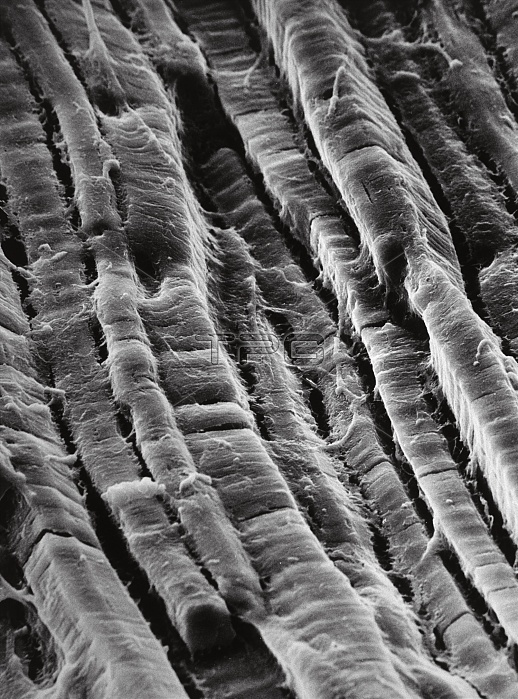
Scanning electron micrograph (SEM) of the surface of a piece of cooked roast beef (bought from a supermarket). The picture shows the parallel muscle fibres, a typical feature of mammalian skeletal muscle. Details of striations - the horizontal banding along each fibre - can also be made out. These striations correspond to the arrangement of actin & myosin filaments in the living muscle. The entire specimen is coated with a thin layer of fat released by the cooking. Magnification: x57 at 35mm size, x400 at 8x10-inch size. Reference: IMICROCOSMOS i, figure 9.19, page 184.
| px | px | dpi | = | cm | x | cm | = | MB |
Details
Creative#:
TOP11722087
Source:
達志影像
Authorization Type:
RM
Release Information:
須由TPG 完整授權
Model Release:
NO
Property Release:
NO
Right to Privacy:
No
Same folder images:
 Loading