
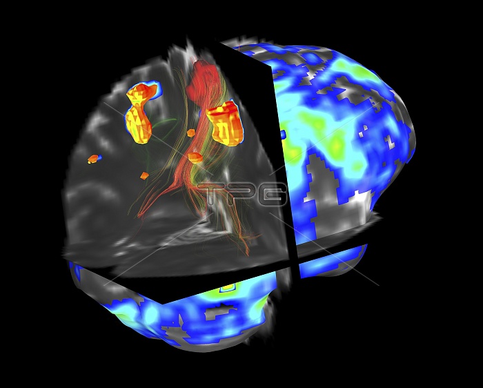
Brain tumour, fMRI and tractography. Combined functional magnetic resonance imaging (fMRI, blue and green) and tractography (yellow and red) imaging of a brain with a tumour (upper left). The front of the brain is at right. The tumour (solid red) has been imaged using tractography, also known as 3D diffusion tensor imaging (DTI) magnetic resonance imaging (MRI). It shows the nerve pathways (red and yellow) affected by the tumour. Brain tumours can be benign or malignant (cancerous). They can cause seizures, headaches, and memory and personality changes. Hot spots on the fMRI areas are activated functional areas.
| px | px | dpi | = | cm | x | cm | = | MB |
Details
Creative#:
TOP11721142
Source:
達志影像
Authorization Type:
RM
Release Information:
須由TPG 完整授權
Model Release:
NO
Property Release:
NO
Right to Privacy:
No
Same folder images:
 Loading