
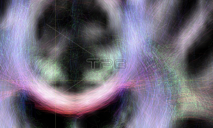
Brain's white matter. Close-up of an area of the brain imaged using tract density imaging and 3D diffusion tensor imaging (DTI), a magnetic resonance imaging (MRI) technique. This frontal view of the centrum semiovale (white matter area) shows crossing areas between the corpus callosum (red), the corticospinal tract (blue) and the superior longitudinal fasciculus (green). The fibres are locally coloured red-green-blue if they are orientated in x-y-z alignment (left-right, posterior-anterior, inferior-superior). Diffusion tensor imaging (tractography) measures the direction of water diffusion, which in the brain reveals the orientation of nerve fibres.
| px | px | dpi | = | cm | x | cm | = | MB |
Details
Creative#:
TOP11721080
Source:
達志影像
Authorization Type:
RM
Release Information:
須由TPG 完整授權
Model Release:
NO
Property Release:
NO
Right to Privacy:
No
Same folder images:
 Loading