
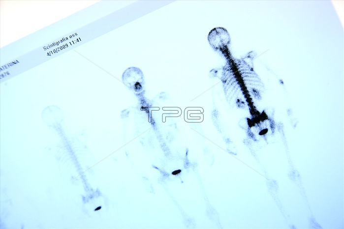
Planar total body scintigraphy of a patient. Scintigraphy is a diagnostic test in which a two dimensional picture of a body radiation source is obtained through the use of radioisotopes. Bone scintigraphy is an important modality for examination of skeletal pathology. Non osseous structures normally seen on bone scintigrams are kidneys and bladder, as technetium 99m diphosphonates are excreted primarily via the urinary tract. it is used occasionally for the evaluation of extra skeletal abnormalities such as myositis ossificans, electrical burns, polymyositis and dermatomyositis, in most cases important information regarding the soft tissues are an incidental finding. The mechanisms of uptake in extra skeletal are similar to those for bones. Calcium and phosphate are deposited when there is an excess of tissue calcium, following local tissue necrosis or damage, or when the calcium phosphate product is elevated such as in primary or secondary hyperparathyroidism. Other mechanisms suggested to explain uptake in tissues other than bone include hyperemia, altered capillary permeability, presence of iron deposits, adsorption onto immature collagen, binding to denatured proteins or enzyme receptors.A radiotracer (bone seeking radionuclide) is injected into the bloodstream through a vein. As it decays, the radiotracer emits gamma radiation, which is detected by a camera that slowly scans your body. The camera captures images of how much radiotracer collects in the bones.
| px | px | dpi | = | cm | x | cm | = | MB |
Details
Creative#:
TOP11657092
Source:
達志影像
Authorization Type:
RM
Release Information:
須由TPG 完整授權
Model Release:
No
Property Release:
No
Right to Privacy:
No
Same folder images:
 Loading