
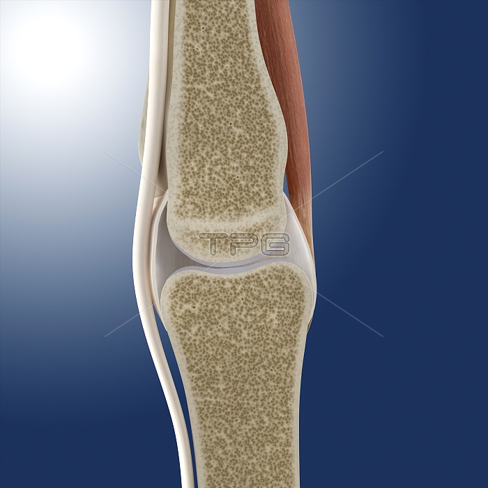
Phalanx joint anatomy. Artwork of a sectioned joint between two phalanx bones (phalanges). These are found in the fingers and toes. The sectioning shows the cancellous interior of the bones, with denser cortical bone forming the outer layers. Cartilage covers the end of the bones where they articulate to form the joint. Ligaments, tendons and other connective tissue (white) hold the joint together and connect bone to bone and muscle (red) to bone.
| px | px | dpi | = | cm | x | cm | = | MB |
Details
Creative#:
TOP11246522
Source:
達志影像
Authorization Type:
RM
Release Information:
須由TPG 完整授權
Model Release:
No
Property Release:
No
Right to Privacy:
No
Same folder images:
PHALANXBONEJOINTTENDONLIGAMENTHANDFOOTFINGERTOEBIOLOGYANATOMYOSTEOLOGYARTHROLOGYARTWORKILLUSTRATIONHYALINECARTILAGECORTICALBONEINTERPHALANGEALARTICULATIONOSSEOUSTISSUECANCELLOUSBONECARTILAGEPHALANXBONETENDONSHEATHDIGITNORMALHEALTHYSECTIONSECTIONEDBLUEBACKGROUNDPHALANGESSPONGYBONECONNECTIVETISSUEJOINTCAPSULEARTICULATINGARTICULATIONMUSCLEBIOLOGICALANATOMICAL
ANATOMICALANATOMYARTHROLOGYARTICULATINGARTICULATIONARTICULATIONARTWORKBACKGROUNDBIOLOGICALBIOLOGYBLUEBONEBONEBONEBONEBONECANCELLOUSCAPSULECARTILAGECARTILAGECONNECTIVECORTICALDIGITFINGERFOOTHANDHEALTHYHYALINEILLUSTRATIONINTERPHALANGEALJOINTJOINTLIGAMENTMUSCLENORMALOSSEOUSOSTEOLOGYPHALANGESPHALANXPHALANXSECTIONSECTIONEDSHEATHSPONGYTENDONTENDONTISSUETISSUETOE
 Loading