
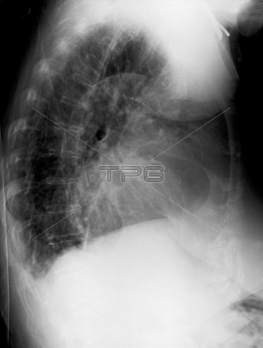
Chest x-ray of a 79 year old woman showing marked cardiomegaly. Diffuse calcifications are observed in the aorta. Pulmonary vascular congestion present suggestive of congestive heart failure (CHF) and pulmonary edema. Increased interstitial densities are seen in the middle and lower lung fields
| px | px | dpi | = | cm | x | cm | = | MB |
Details
Creative#:
TOP10757603
Source:
達志影像
Authorization Type:
RM
Release Information:
須由TPG 完整授權
Model Release:
No
Property Release:
No
Right to Privacy:
No
Same folder images:
technologydistorteddiagnosisresearchradiologyproblemsphotographyblackandwhitesciencecongestionverticalbiologychestclose-upenlargedbonex-rayimaginganatomydiagnosticaortarespiratorysystempulmonaryblackbackgroundmedicalsciencemisfortunefailurelimbscrutinyheartdiseasehumanheartanatomicalhumanskeletonAtherosclerosisHumanLungHumanInternalOrganHumanBoneHealthcareAndMedicineHumanBodyPartMedicalExammedicalexaminationradiographynopeopleRadiogramX-RayImageScientificImagingTechniqueCalcificationradiographcardiomegalyEdemaDiagnosticAidCardiovascularDiseasecongestiveCHFBoneSystemCuboidBoneMedialImageryScientificImagery
AidAndAtherosclerosisBodyBoneBoneBoneCHFCalcificationCardiovascularCuboidDiagnosticDiseaseEdemaExamHealthcareHumanHumanHumanHumanImageImageryImageryImagingInternalLungMedialMedicalMedicineOrganPartRadiogramScientificScientificSystemTechniqueX-Rayanatomicalanatomyandaortabackgroundbiologyblackblackbonecardiomegalychestclose-upcongestioncongestivediagnosisdiagnosticdiseasedistortedenlargedexaminationfailurehearthearthumanhumanimaginglimbmedicalmedicalmisfortunenopeoplephotographyproblemspulmonaryradiographradiographyradiologyresearchrespiratorysciencesciencescrutinyskeletonsystemtechnologyverticalwhitex-ray
 Loading