
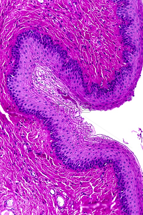
Stratified squamous epithelium. Light micrograph of a section through stratified squamous epithelium, showing the multiple layers, or strata, that function to resist abrasion of the surface. This is the epithelial pattern found within the epidermis of the skin, but in this image, the absence of a layer of keratin is typical of the oesophagus (gullet). Squamous cells on the surface are continuously lost by cell death and exfoliation, but are replaced by the proliferation of stem cells located at the base of the epithelium. New epithelial cells mature and slowly displace towards the surface. The connective tissue beneath the epithelium, the lamina propria, contains nerves and blood vessels. Magnification: x139, when printed 10 centimetres
| px | px | dpi | = | cm | x | cm | = | MB |
Details
Creative#:
TOP10709103
Source:
達志影像
Authorization Type:
RM
Release Information:
須由TPG 完整授權
Model Release:
No
Property Release:
No
Right to Privacy:
No
Same folder images:
 Loading