
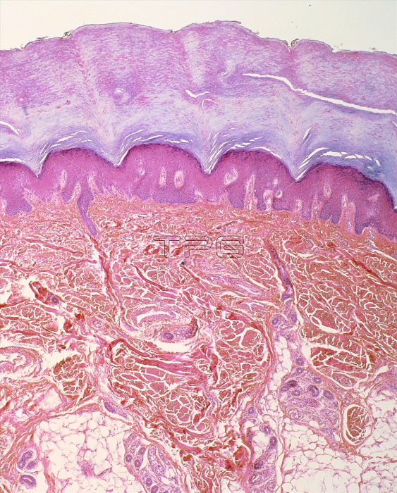
Light micrograph of a section through the skin of the finger. Four principal layers are discernable here: the outer epidermis (mauve), composed of dead, flattened, cornified cells; the lower living part of the epidermis (pink); the dermis (red), composed of dense, fibro-elastic tissue threaded with blood vessels; and a layer of subcutaneous fat (white). The purple-stained regions lying at the base of the dermis are sweat glands. Each one is an unbranched, tightly-coiled tube, linked to the skin's surface by a sweat duct. Part of a duct is visible extending upwards from the sweat gland at lower left. Magnification: x25 at 6x7cm size.
| px | px | dpi | = | cm | x | cm | = | MB |
Details
Creative#:
TOP10222998
Source:
達志影像
Authorization Type:
RM
Release Information:
須由TPG 完整授權
Model Release:
N/A
Property Release:
N/A
Right to Privacy:
No
Same folder images:
 Loading