
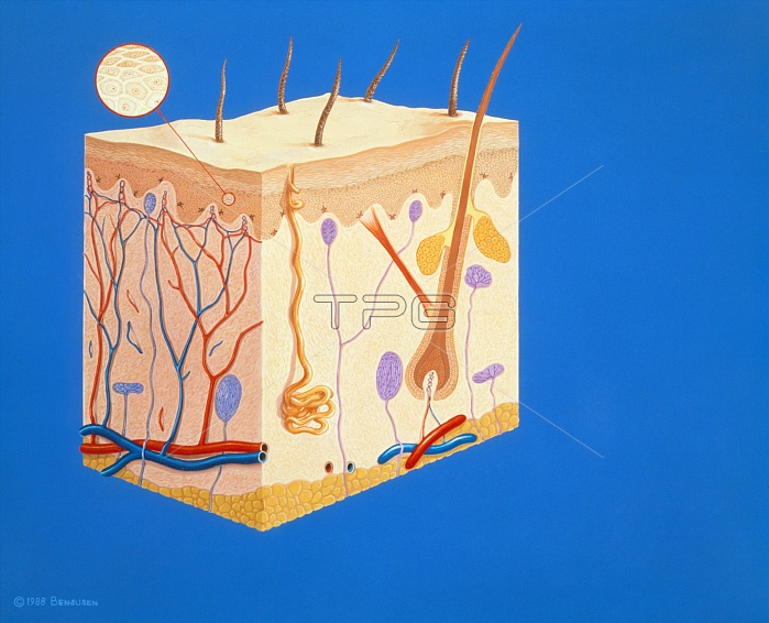
Illustration showing a cross-section through the human skin. The outer layer is the epidermis, a protective layer of flattened, horny cells impregnated with keratin (inset). Below this are the dermis (a thick layer of connective tissue) & a layer of subcutaneous fat. Within the dermis are blood capillaries (red & blue), sensory nerve endings (purple), an eccrine sweat gland (beige) & a hair follicle. Attached to the hair follicle are sebaceous glands (yellow) and the hair erector muscle. Four types of nerve endings are shown: Meissner's corpuscles (top), Pacinian corpuscles (large ovals), Ruffini's corpuscles (eg. bottom right) and a Krause's end bulb (centre right).
| px | px | dpi | = | cm | x | cm | = | MB |
Details
Creative#:
TOP10222990
Source:
達志影像
Authorization Type:
RM
Release Information:
須由TPG 完整授權
Model Release:
N/A
Property Release:
N/A
Right to Privacy:
No
Same folder images:
 Loading