
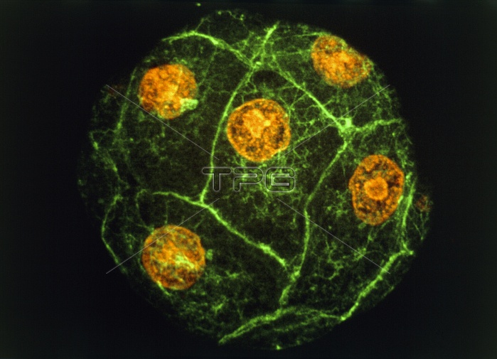
Sea urchin embryo. Immunofluorescence micrograph of a sea urchin embryo at the 8-16 cell stage. The orange structures are the cell nuclei. The bound- aries between different cells show up as green lines. Following fertilization an embryo undergoes multiple rounds cell division, dividing each time into double the number of cells (1, 2, 4, 8 ...). Eventually a hollow ball of hundreds of cells is produced, which then starts to differentiate. This picture was made by exposing the embryo to fluor- escent antibodies that bind to certain proteins in the cell. The embryo was then viewed with a laser- scanning light microscope, which makes the anti- bodies fluoresce. Magnification unknown.
| px | px | dpi | = | cm | x | cm | = | MB |
Details
Creative#:
TOP10222538
Source:
達志影像
Authorization Type:
RM
Release Information:
須由TPG 完整授權
Model Release:
N/A
Property Release:
N/A
Right to Privacy:
No
Same folder images:
 Loading