
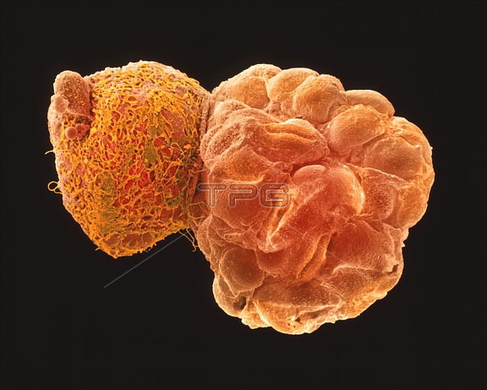
Hatching blastocyst embryo. Coloured scanning electron micrograph (SEM) of a human embryo at the blastocyst stage, five days after fertilisation. It is seen at right hatching from a hole in the zona pellucida (at left), a protein shell that originally surrounded the unfertilised egg. The blastocyst is a hollow ball of cells with a fluid centre. Each cell is called a blastomere. Most of these embryo cells will form the placenta and membranes around the embryo, and only a small group (the inner mass) form the embryo proper. At this stage the blastocyst has moved into the uterus (womb) and is preparing to implant on the womb wall. Magnification: x300 at 6x7cm size. Mag;x290 at original 6x7cm size.
| px | px | dpi | = | cm | x | cm | = | MB |
Details
Creative#:
TOP10222223
Source:
達志影像
Authorization Type:
RM
Release Information:
須由TPG 完整授權
Model Release:
N/A
Property Release:
N/A
Right to Privacy:
No
Same folder images:
 Loading