
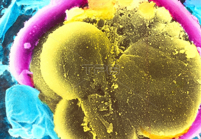
4-6 cell embryo. Coloured scanning electron micrograph (SEM) of layers of a 4-6 cell embryo. The blastomeres (large, yellow) are the cells formed from divisions of the fertilized egg (ovum). Smaller fragments" of blastomeres are evident at centre top. Surrounding the embryo is a membranous envelope, the zona pellucida (pink). Beyond the zona pellucida is a layer composed of the metabolically active cumulus cells (blue). The cumulus cells provide the correct microenvironment for fertilization. Sperm must penetrate this cell layer in order to fertilize the egg. Magnification: x2,600 at 5x7cm size."
| px | px | dpi | = | cm | x | cm | = | MB |
Details
Creative#:
TOP10222211
Source:
達志影像
Authorization Type:
RM
Release Information:
須由TPG 完整授權
Model Release:
N/A
Property Release:
N/A
Right to Privacy:
No
Same folder images:
 Loading