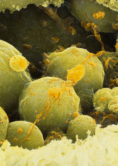
Aborting embryo cells. Coloured scanning electron micrograph (SEM) of blastomere fragments of an aborting 6-8 cell human embryo. The embryo is undergoing necrosis (tissue death). Blastomeres, the cells formed from divisions of the fertilized egg, have degenerated into fragments (green). Remains of sperm tails (orange) can also be seen. The zona pellucida (bottom, yellow), the membrane which surrounds the embryo, has been artificially fractured (opened up). It has become thick and compact and has lost its usual striated appearance. Magnification: x3,800 at 5x7cm size.
| px | px | dpi | = | cm | x | cm | = | MB |
Details
Creative#:
TOP10222210
Source:
達志影像
Authorization Type:
RM
Release Information:
須由TPG 完整授權
Model Release:
N/A
Property Release:
N/A
Right to Privacy:
No
Same folder images:

 Loading