
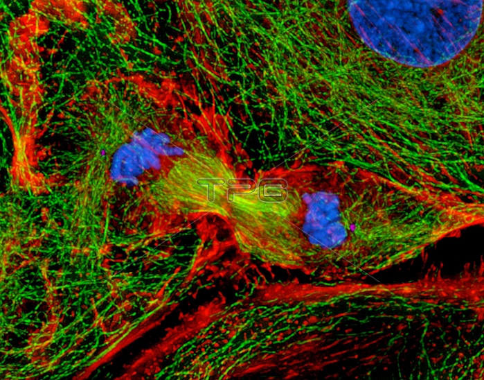
Mitosis. Digital three-dimensional immunofluor- escent light micrograph of a section through a rat kangaroo kidney epithelial cell during cytokin- esis, cytoplasmic cleavage to form two cells. This is the telophase stage of mitotic cell division, and nuclear membranes (not seen) are forming around the two groups of chromosomes (blue, upper left & centre right). The pinched cytoplasm is at centre. The actin microfilaments (red) and tubulin microtubules (green) of the cytoskeleton maintain the structure of the cell. Mitosis produces two identical daughter cells. Antibodies have been used to attach fluorescent dyes to specific cell tissues. Magnification: x500 at 6x7cm size.
| px | px | dpi | = | cm | x | cm | = | MB |
Details
Creative#:
TOP10222114
Source:
達志影像
Authorization Type:
RM
Release Information:
須由TPG 完整授權
Model Release:
N/A
Property Release:
N/A
Right to Privacy:
No
Same folder images:
 Loading