
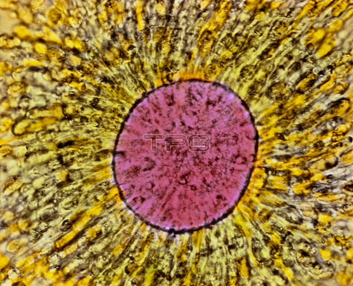
Mature oocyte. Coloured light micrograph of a human secondary oocyte (mature egg). At centre is the rounded egg (pink) as seen in the ovary before ovulation. The egg cell undergoes meiotic division giving rise to two cells: a secondary oocyte and a polar body (not seen). The polar body degenerates and has no function. Here, directly around the egg is the zona pellucida single layer of cells (unfocused); outside the zona pellucida are several layers of cells making up the corona radiata (yellow). These cells support and nourish the developing egg in the ovary. Magnification: x560 at 6x7cm size. x740 at 4x5"~LANDSCAPE"
| px | px | dpi | = | cm | x | cm | = | MB |
Details
Creative#:
TOP10221683
Source:
達志影像
Authorization Type:
RM
Release Information:
須由TPG 完整授權
Model Release:
N/A
Property Release:
N/A
Right to Privacy:
No
Same folder images:
 Loading