
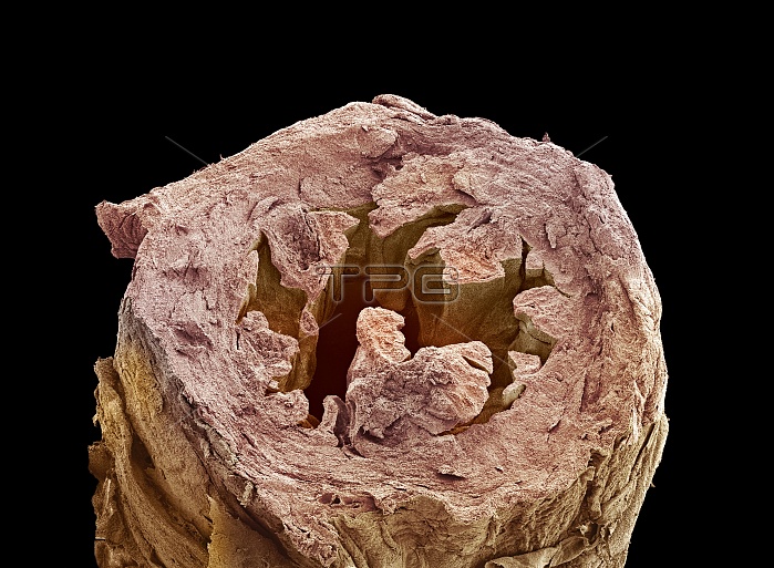
Fallopian tube. Coloured scanning electron micrograph (SEM) of a cross-section through a fallopian tube (oviduct). The fallopian tubes carry the egg from the ovary to the uterus (womb). The interior (lumen) of the tube is lined with a folded mucous membrane, which is surrounded by a thin layer of muscle. The outer layer, the serosa, consists of connective tissue and is highly vascular. Magnification: x11 when printed 10 centimetres wide.
| px | px | dpi | = | cm | x | cm | = | MB |
Details
Creative#:
TOP10221515
Source:
達志影像
Authorization Type:
RM
Release Information:
須由TPG 完整授權
Model Release:
N/A
Property Release:
N/A
Right to Privacy:
No
Same folder images:
FALLOPIANTUBEOVIDUCTHUMANBODYHISTOLOGYBIOLOGYPHYSIOLOGYANATOMYFEMALESEMCOLOUREDSCANNINGELECTRONMICROSCOPELANDSCAPEPHYSIOLOGICALBIOLOGICALFALSE-COLOURSCANNINGELECTRONMICROGRAPHHORIZONTALMUCOUSMEMBRANEEPITHELIUMEPITHELIAEPITHELIALREPRODUCTIONREPRODUCTIVETRACTSYSTEMFOLDEDMUSCLEMUSCULARISSEROSASEROSALCONNECTIVETISSUEHISTOLOGICALCROSS-SECTIONSECTION"
"ANATOMYBIOLOGICALBIOLOGYBODYCOLOUREDCONNECTIVECROSS-SECTIONELECTRONELECTRONEPITHELIAEPITHELIALEPITHELIUMFALLOPIANFALSE-COLOURFEMALEFOLDEDHISTOLOGICALHISTOLOGYHORIZONTALHUMANLANDSCAPEMEMBRANEMICROGRAPHMICROSCOPEMUCOUSMUSCLEMUSCULARISOVIDUCTPHYSIOLOGICALPHYSIOLOGYREPRODUCTIONREPRODUCTIVESCANNINGSCANNINGSECTIONSEMSEROSASEROSALSYSTEMTISSUETRACTTUBE
 Loading