
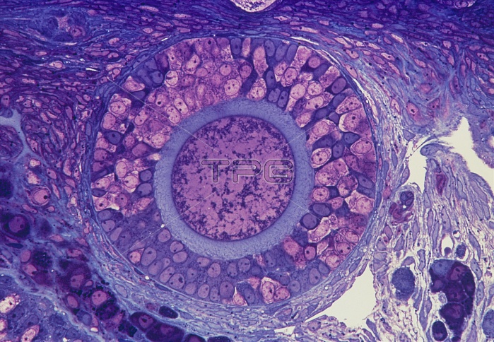
Ovarian follicle. Light micrograph of a sectioned ovary showing a primary follicle (large pink and purple circle). Within the follicle is the primary oocyte (smaller circle, centre) which contains the chromosomes (not seen). This is surrounded by several layers of granulosa cells (pink and purple, outer circle) which secrete glycoproteins. These form the zona pellucida (blue) which separates the oocyte from the granulosae and is penetrated by the sperm at fertilization. Several follicles mature with every ovarian cycle, but only one becomes a Graafian follicle, releasing its oocyte at ovulation. Toluidine blue stain. Magnification: x80 at 35mm size.
| px | px | dpi | = | cm | x | cm | = | MB |
Details
Creative#:
TOP10221453
Source:
達志影像
Authorization Type:
RM
Release Information:
須由TPG 完整授權
Model Release:
N/A
Property Release:
N/A
Right to Privacy:
No
Same folder images:
 Loading