
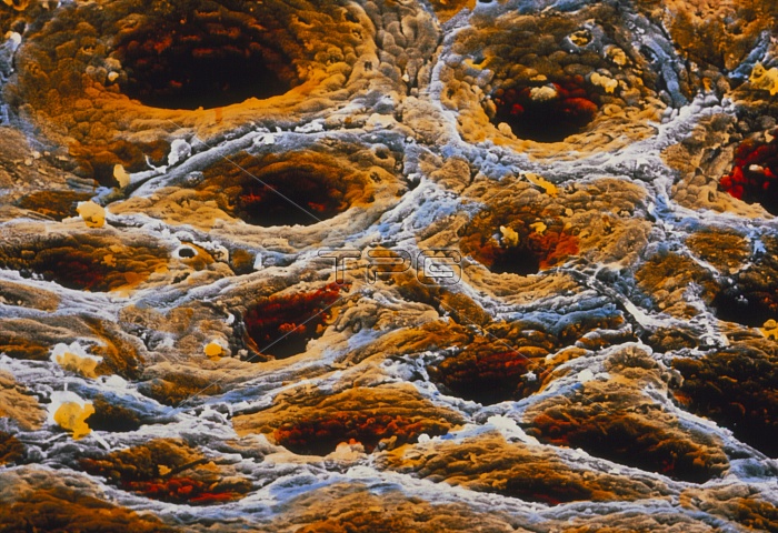
False-colour scanning electron micrograph (SEM) of the glandular wall of the colon. Many open- ings to glands can be seen. Desquamating dead cells are bright yellow. The mucosa of the wall has few folds; here, in the colon, undigestable food becomes faeces with the absorption of excess water. Columnar surface cells (visible here) are involved in this water absorption. Goblet cells largely within the gland openings secrete mucous to protect the colon surface and lubricate faeces. The large intestine is some 1.5 metres long, with regions that include the appendix, colon and rectum. Magnification: x160 at 6x7cm size. Magnification: x240 at 4x5 inch size.
| px | px | dpi | = | cm | x | cm | = | MB |
Details
Creative#:
TOP10220996
Source:
達志影像
Authorization Type:
RM
Release Information:
須由TPG 完整授權
Model Release:
N/A
Property Release:
N/A
Right to Privacy:
No
Same folder images:
 Loading