
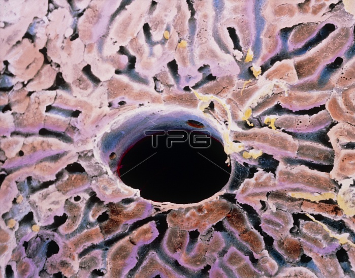
False-colour scanning electron micrograph (SEM) of a lobule of the liver. Parenchyma tissue in the liver is arranged into lobules of cells, one of which is seen here. Each lobule has a central vein (black circle here); portal tracts at the boundary of each lobule (beyond this field of view) lead blood to flow along paths or sinusoids (grey) between hepatic cells, towards the centre of the lobule. In the process, blood is detoxified of waste products; proteins are metabolized; blood clotting agents, vitamin A and bile are synthe- sized; and spent red blood cells are reabsorbed. Magnification: x570 at 6x7cm size. Magnification: x875 at 4x5 inch size.
| px | px | dpi | = | cm | x | cm | = | MB |
Details
Creative#:
TOP10220709
Source:
達志影像
Authorization Type:
RM
Release Information:
須由TPG 完整授權
Model Release:
N/A
Property Release:
N/A
Right to Privacy:
No
Same folder images:
 Loading