
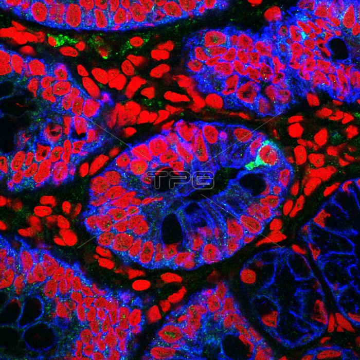
Small intestine. Fluorescence confocal light micrograph of a horizontal section through a human small intestine. The small intestine runs from the stomach to the large intestine. It is where digestion is completed and nutrients are absorbed into the blood. The interior (lumen) is lined with villi (blue amd pink ovals), which are folds in the intestinal surface that greatly increase the surface area for absorption. As well as absorptive cells the villi contain goblet cells, which secrete mucus to lubricate the passage of the partly digested food through the intestine, and neuroendocrine cells (green), which secrete hormones into the bloodstream.
| px | px | dpi | = | cm | x | cm | = | MB |
Details
Creative#:
TOP10220688
Source:
達志影像
Authorization Type:
RM
Release Information:
須由TPG 完整授權
Model Release:
N/A
Property Release:
N/A
Right to Privacy:
No
Same folder images:
 Loading