
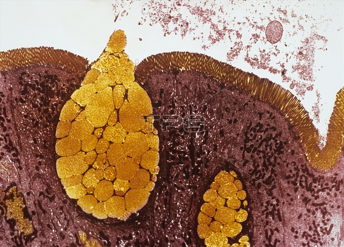
Intestinal lining. Coloured transmission electron micrograph (TEM) of a section through the wall of the small intestine. On the surface of the intestinal wall are numerous microvilli, tiny finger-like projections, that increase the surface area available for the absorption of nutrients. Within the epithelium (purple) are goblet cells (yellow). These cells secrete mucus, which protects the lining of the intestine. Inside the goblets cells are many mucigen granules (round). When these are released into the intestine they will combine with water to form mucin, the main constituent of mucus. Magnification: x2850 when printed at 10 centimetres wide.
| px | px | dpi | = | cm | x | cm | = | MB |
Details
Creative#:
TOP10220667
Source:
達志影像
Authorization Type:
RM
Release Information:
須由TPG 完整授權
Model Release:
N/A
Property Release:
N/A
Right to Privacy:
No
Same folder images:
 Loading