
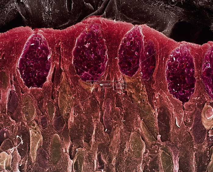
Small intestine villus. Coloured scanning electron micrograph (SEM) of a freeze fracture section through a villus from the mucosal lining of the small intestine. Villi are finger-like projections that increase the surface area of a structure. Microvilli, just visible across upper centre, further increase the surface area available for food absorption. The outer surface of a villus is mostly columnar epithelium (red). It contains numerous goblet cells (dark pink), which secrete mucus to lubricate food & prevent self-digestion. Within the goblet cells individual mucin granules are seen. Magnification: x2000 at 6x7cm size.
| px | px | dpi | = | cm | x | cm | = | MB |
Details
Creative#:
TOP10220615
Source:
達志影像
Authorization Type:
RM
Release Information:
須由TPG 完整授權
Model Release:
N/A
Property Release:
N/A
Right to Privacy:
No
Same folder images:
 Loading