
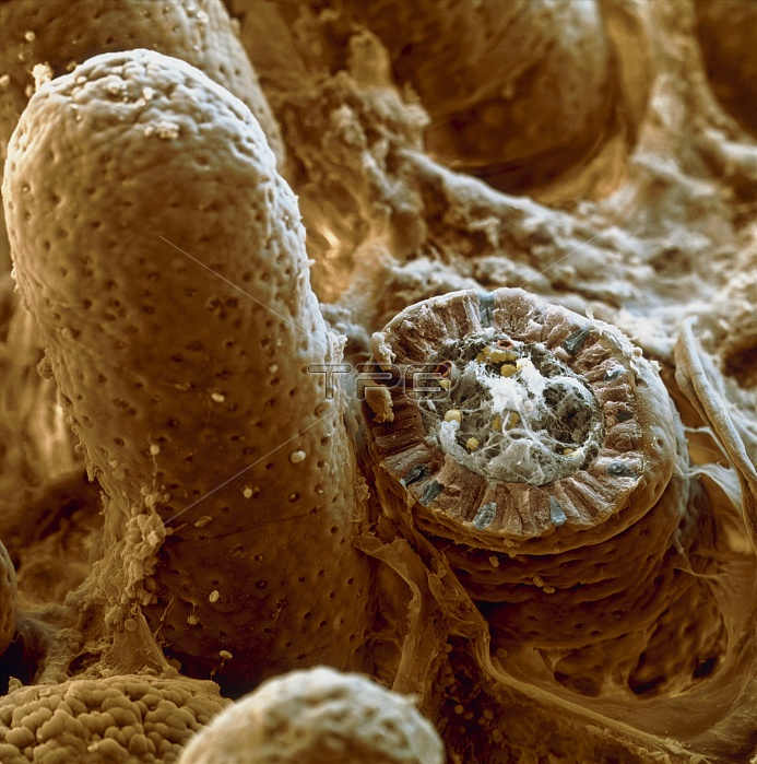
Small intestine villi. Coloured scanning electron micrograph (SEM) of villi (brown) on the lining of the small intestine. Villi greatly increase the intestinal surface area for the absorption of nutrients from food. A broken villus is seen at lower right, revealing its internal structure. The villus epithelium (brown/blue in cross- section) contains enterocytes (light brown), which are involved in nutrient absorption. Scattered amongst these are goblet cells (blue), which secrete mucus onto the intestinal surface. Capillary blood vessels (red) within the villus transport digestive products to a nearby vein. Magnification: x190 at 6x6cm size. x300 at 4x5"~SQUARE"
| px | px | dpi | = | cm | x | cm | = | MB |
Details
Creative#:
TOP10220609
Source:
達志影像
Authorization Type:
RM
Release Information:
須由TPG 完整授權
Model Release:
N/A
Property Release:
N/A
Right to Privacy:
No
Same folder images:
 Loading