
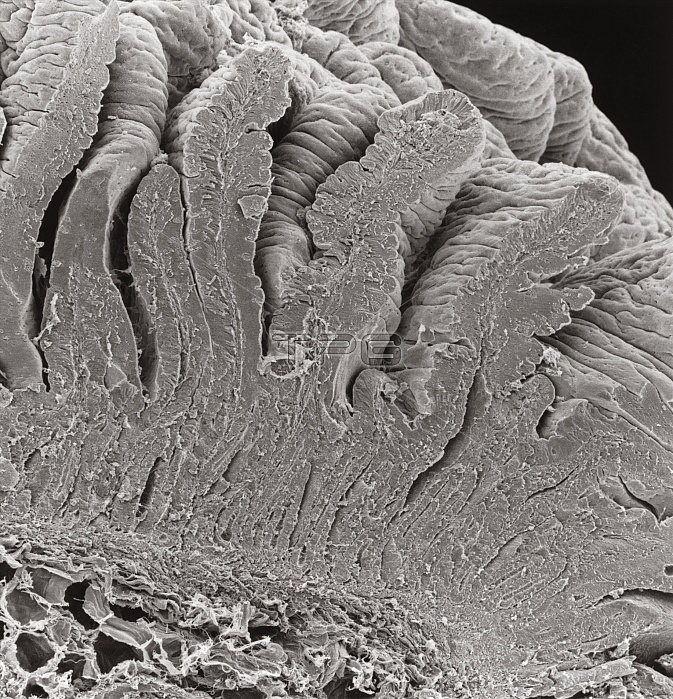
Scanning electron micrograph of a section through the wall of the human duodenum, showing the many tiny folds, known as villi, which project 0.5 to 1 mm out into the intestinal lumen. These folds greatly increase the effective absorptive and secretory surface of the mucosa (mucus membrane) which lines the small intestine. Each villus contains a central core of connective tissue, known as the lamina propria (just visible as a strand of tissue running up the centre of the cut ends). This contains large blood vessels, capillaries, some smooth muscle cells and a blind- ended lymph vessel known as a lacteal. Magnification: x220 at 8x10 inch size.
| px | px | dpi | = | cm | x | cm | = | MB |
Details
Creative#:
TOP10220570
Source:
達志影像
Authorization Type:
RM
Release Information:
須由TPG 完整授權
Model Release:
N/A
Property Release:
N/A
Right to Privacy:
No
Same folder images:
 Loading