
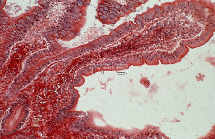
Light micrograph of villi in the normal human small intestine. Villi, finger-like projections into the lumen of the small intestine, are a modification designed to increase the absorptive area of the surface. The core of the villus consists of a connective tissue called the lamina propria, & smooth muscle, stained red. The surface of the villus is bounded by epithelial cells and goblet cells, seen as large, roundish objects with granular purple contents. The absorptive epithelial cells have microvilli packed on their outer edge - a thin dark red line next to the unstained lumen. Magnification: x50 at 35mm size.
| px | px | dpi | = | cm | x | cm | = | MB |
Details
Creative#:
TOP10220565
Source:
達志影像
Authorization Type:
RM
Release Information:
須由TPG 完整授權
Model Release:
N/A
Property Release:
N/A
Right to Privacy:
No
Same folder images:
 Loading