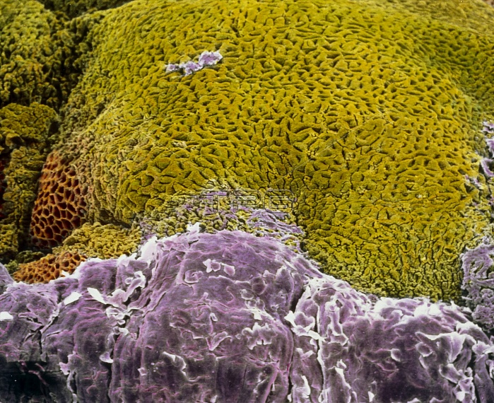
False-colour scanning electron micrograph (SEM) of the transition zone between the oesophagus and stomach. Below, the stratified squamous epithelium of the oesophagus (violet), contrasts with the glandular wall of the stomach mucosa (green). The surface of the oesophagus is thick, multi-layered, and adapted for the movement of food along it. While the stomach mucosa shows many orifices of glands which secrete digestive enzymes, hydrochloric acid, and hormones; a simple columnar epithelium secretes mucous. At left (brown), part of the stomach wall is destroyed in an ulcer-like manner. Magnification: x90 at 6x7cm size. Magnification: x140 at 4x5 inch size.
| px | px | dpi | = | cm | x | cm | = | MB |
Details
Creative#:
TOP10220492
Source:
達志影像
Authorization Type:
RM
Release Information:
須由TPG 完整授權
Model Release:
N/A
Property Release:
N/A
Right to Privacy:
No
Same folder images:

 Loading