
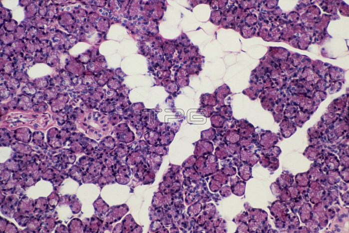
Light micrograph of normal human parotid gland. One of the salivary glands, the parotid consists of acini arranged in lobes. This picture shows a junction between several lobes; the clear spaces represent the interlobular connective tissue. The masses of secretory cells (granular pink, purple nuclei) produce a serous (watery) secretion which is passed to intralobular ducts. Three are visible here; two centre left and one bottom right. In cross section (two here) the ducts appear ring shaped with a central space surrounded by peripheral rows of cells (clear pink with purple nuclei). Magnification: x50 at 35mm size.
| px | px | dpi | = | cm | x | cm | = | MB |
Details
Creative#:
TOP10220369
Source:
達志影像
Authorization Type:
RM
Release Information:
須由TPG 完整授權
Model Release:
N/A
Property Release:
N/A
Right to Privacy:
No
Same folder images:
 Loading