
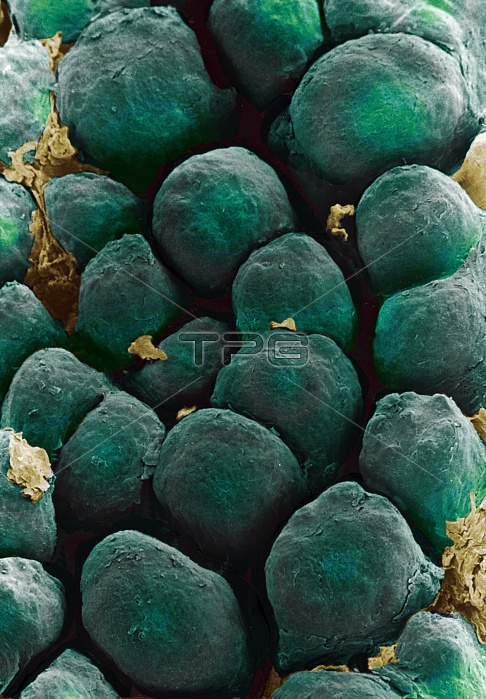
Tongue papillae. Coloured scanning electron micrograph (SEM) of filiform papillae on the surface of the tongue. Filiform papillae are covered by stratified squamous epithelial cells. Dead cells of the uppermost layer are constantly being shed and replaced (desquamation). This shedding gives the papillae their scaly appearance. Filiform papillae form a rough surface to aid chewing. Each papilla contains nerve endings which transmit tactile (touch) information to the brain. Magnification unknown.
| px | px | dpi | = | cm | x | cm | = | MB |
Details
Creative#:
TOP10220348
Source:
達志影像
Authorization Type:
RM
Release Information:
須由TPG 完整授權
Model Release:
N/A
Property Release:
N/A
Right to Privacy:
No
Same folder images:
 Loading