
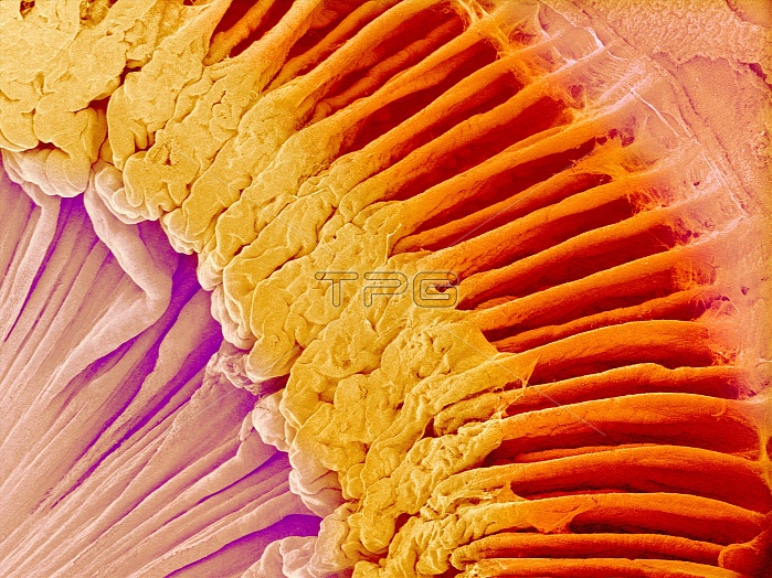
Eye anatomy. Coloured scanning electron micrograph (SEM) of part of the ciliary body (red/yellow) of the eye, a ring-shaped structure inside the eye, surrounding the iris. This view looks at part of the ciliary body (arching from upper left to lower right), as seen from inside the eye. Part of the iris is seen at lower left. Part of the choroid (outer layer of the eye) is at top right. The ciliary body joins to ligaments that hold the lens in place behind the iris. The lens has been removed here. The ciliary body also contains the ciliary muscle that is contracted to alter the curvature of the lens and focus light on the retina at the back of the eye. These structures are at the front of the eye.
| px | px | dpi | = | cm | x | cm | = | MB |
Details
Creative#:
TOP10220091
Source:
達志影像
Authorization Type:
RM
Release Information:
須由TPG 完整授權
Model Release:
N/A
Property Release:
N/A
Right to Privacy:
No
Same folder images:
 Loading