
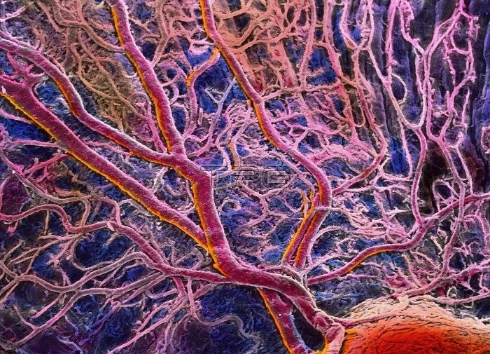
False-colour scanning electron micrograph (SEM) of blood vessels in the choroid of the eye. A branch- ing network of arteries and veins can be seen in this area under the central fovea. The choroid is a tissue layer beneath the retina which supplies blood, thus food and oxygen, to light sensitive cells of the eye. Many cone cells occur on the retina at the central fovea area, specialized for acute day vision, and hence an area that uses much blood. The choroid is also darkened (blue) with pigment cells; pigment absorbs light rays passing through the retina and prevents light reflection. Magnification: x46 at 6x7cm size. Magnification: x70 at 4x5 inch size.
| px | px | dpi | = | cm | x | cm | = | MB |
Details
Creative#:
TOP10220015
Source:
達志影像
Authorization Type:
RM
Release Information:
須由TPG 完整授權
Model Release:
N/A
Property Release:
N/A
Right to Privacy:
No
Same folder images:
 Loading