
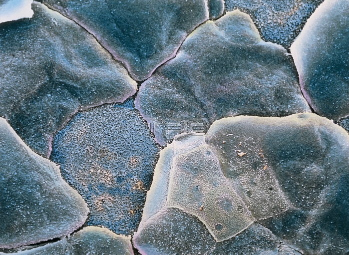
False-colour scanning electron micrograph (SEM) of the cell surface of the cornea of the eye. These cells are flattened, closely packed, and trans- parent. This is a stratified squamous epithelium 5 cell layers deep. Cells at centre left and upper right have peeled away, to reveal tiny ridges (microplicae) on the inner surface which bind each cell together. Although rich in nerves, there are no blood vessels in the cornea: this would affect its transparency. This external surface over the anterior of the eye has a convex curve to provide the main mechanism for focusing light onto the retina. Magnification: x1170 at 6x7cm size. Magnification: x1800 at 4x5 inch size.
| px | px | dpi | = | cm | x | cm | = | MB |
Details
Creative#:
TOP10220014
Source:
達志影像
Authorization Type:
RM
Release Information:
須由TPG 完整授權
Model Release:
N/A
Property Release:
N/A
Right to Privacy:
No
Same folder images:
 Loading