
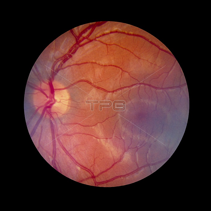
Fundus camera image of the retina of a normal eye, showing the distribution of the retinal veins & arteries: the central retinal artery (a branch of the opthalmic artery) enters the optic nerve 2 cm before it reaches the eyeball, emerging through the retina in the centre of the optic disc (the blind spot of the eye, the pale area on the left). The dark oval region free of large blood vessels (right) is the macula, in the centre of which the fovea is found. The fovea is the region with highest visual acuity. An extension of the optic nerve, the retina consists of photosensitive cells (rods & cones) which translate light energy into nervous impulses.
| px | px | dpi | = | cm | x | cm | = | MB |
Details
Creative#:
TOP10219995
Source:
達志影像
Authorization Type:
RM
Release Information:
須由TPG 完整授權
Model Release:
N/A
Property Release:
N/A
Right to Privacy:
No
Same folder images:
 Loading