
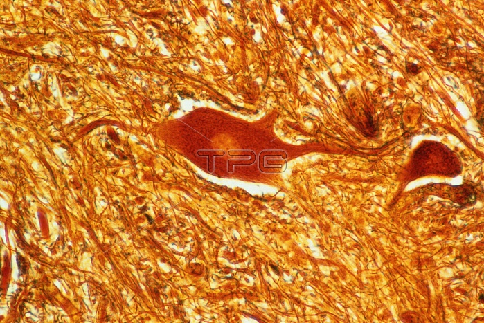
Light micrograph of a motorneuron cell from the ventral horn of a human spinal cord, shown with Bodian staining. The pale central area is the nucleus, surrounded by the brown cell soma and extended dendrites. The soma and dendrites are the main recipient surfaces of the cell. The ventral horn of the spinal column is composed mainly of grey matter, in which motoneurons are found among large areas of connective tissue. Magnification x250 at 5x7cm, x125 at 35mm size.
| px | px | dpi | = | cm | x | cm | = | MB |
Details
Creative#:
TOP10219369
Source:
達志影像
Authorization Type:
RM
Release Information:
須由TPG 完整授權
Model Release:
N/A
Property Release:
N/A
Right to Privacy:
No
Same folder images:
 Loading