
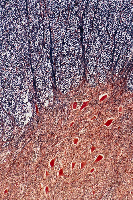
Light micrograph of normal human spinal cord. This cross section shows the junction between the grey matter (bottom, orange) and the white matter (top) in the anterior horn. The large cells, red, (some with pale nucleus and dense nucleolus) are anterior horn cells, whose axons become the efferent root fibres. They are embedded in a mass of neuroglial cells. In cross section (most of upper field) they appear as a clear ring (myelin) around a central dot (the fibres). The prominent ascending lines are fibres cut longitudinally. Magnification: X 20 at 35mm size.
| px | px | dpi | = | cm | x | cm | = | MB |
Details
Creative#:
TOP10219364
Source:
達志影像
Authorization Type:
RM
Release Information:
須由TPG 完整授權
Model Release:
N/A
Property Release:
N/A
Right to Privacy:
No
Same folder images:
 Loading