
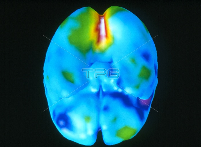
Brain's visual centre. Coloured three-dimensional positron emission tomography (PET) scan of the brain seen from below during visual activity. The frontal lobe is at lower centre. The scan shows the amount of glucose (sugar) consumption and hence areas of low (blue) and high (red & white) brain activity. The most active area is the visual cortex within the occipital lobe at the back of the brain (at upper centre). The cerebellum has been removed to allow this area to be viewed more clearly. PET scans use radioactively-labelled substances introduced into the blood to view metabolic activity in three-dimensions.
| px | px | dpi | = | cm | x | cm | = | MB |
Details
Creative#:
TOP10219327
Source:
達志影像
Authorization Type:
RM
Release Information:
須由TPG 完整授權
Model Release:
N/A
Property Release:
N/A
Right to Privacy:
No
Same folder images:
 Loading