
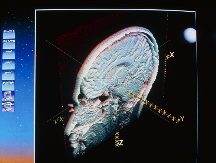
MEG brain signal. Computer image of a section through the human head (sagittal section), showing a region in the brain at which electrical stimulation has occurred. The region is demarcated in three dimensions with axes X, Y and Z. These axes are a method of representing direction and intensity of an electrical impulse in the brain in response to a stimulus. The impulse was detected using magnetoencephalography (MEG), a brain scanning technique that measures magnetic fields generated from nerve cell activity in the brain. The image of the head was contructed from 3-D MRI scan data. Photographed at New York University Medical Centre in New York City, USA.
| px | px | dpi | = | cm | x | cm | = | MB |
Details
Creative#:
TOP10219311
Source:
達志影像
Authorization Type:
RM
Release Information:
須由TPG 完整授權
Model Release:
N/A
Property Release:
N/A
Right to Privacy:
No
Same folder images:
 Loading