
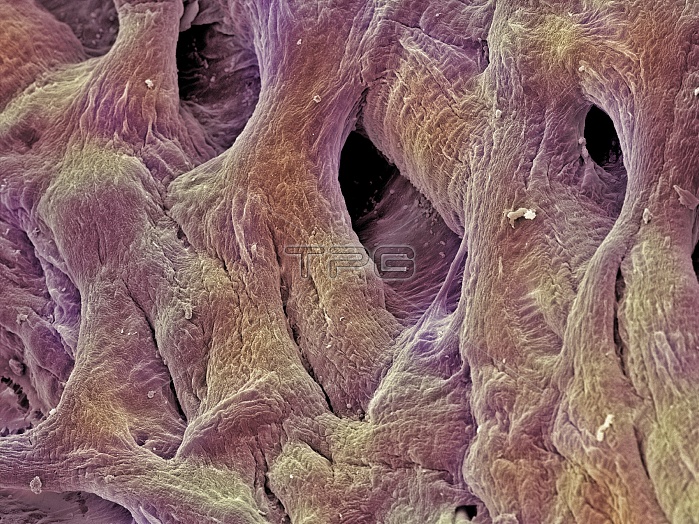
Heart valve tendons. Coloured scanning electron micrograph (SEM) of the chordae tendineae, tendons which attach the atrioventricular heart valves to their muscles. These valves prevent blood being forced back into the atria (upper chambers) from the ventricles (lower chambers) during the contraction of the ventricles. The right ventricle (which pumps deoxygenated blood to the lungs) is separated from the right atrium by the tricuspid valve. The left ventricle (which pumps newly- oxygenated blood around the body) is separated from the left atrium by the mitral valve. The valves are controlled by papillary muscles (not seen). Magnification unknown.
| px | px | dpi | = | cm | x | cm | = | MB |
Details
Creative#:
TOP10218072
Source:
達志影像
Authorization Type:
RM
Release Information:
須由TPG 完整授權
Model Release:
N/A
Property Release:
N/A
Right to Privacy:
No
Same folder images:
 Loading