
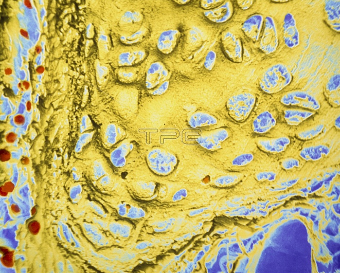
False-colour scanning electron micrograph (SEM) of human hyaline cartilage from the walls of a bronchus, the main conducting airway of a lung. Hyaline cartilage is found in the walls of conducting airways (the trachea & bronchi) and a specialized type of hyaline cartilage caps the ends of long bones that form joints with other bones. This articular cartilage has a smooth slippery surface which combines with the synovial fluid to ease movement in the joint. This image shows the organization of chondrocytes (cartilage cells) which are embedded in a cartilage matrix that the cells have synthesized. Magnification: x220 at 35mm size.
| px | px | dpi | = | cm | x | cm | = | MB |
Details
Creative#:
TOP10217621
Source:
達志影像
Authorization Type:
RM
Release Information:
須由TPG 完整授權
Model Release:
N/A
Property Release:
N/A
Right to Privacy:
No
Same folder images:
 Loading