
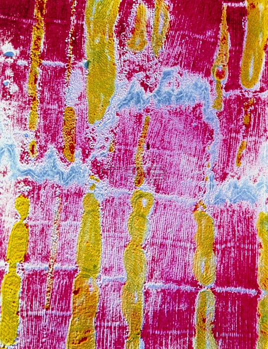
False-colour transmission electron micrograph of a longitudinal section through striated cardiac muscle. Cardiac muscle, unlike skeletal muscle, is composed of seperate cellular elements. The meandering, thick blue line marks the boundary between two muscle cells and is known as a fascia adherens. Each cell is composed of a number of myofibrils (pink), surrounded by sarcoplasm (yellow). The myofibrils are, in turn, composed of contractile proteins arranged in thin threads called myofilaments (visible here as fine, pink, vertical stripes). The presence of two different types of protein causes the pink & blue horizontal banding. Mag: x11,000 at 6x7cm, x5500 at 35mm size
| px | px | dpi | = | cm | x | cm | = | MB |
Details
Creative#:
TOP10217474
Source:
達志影像
Authorization Type:
RM
Release Information:
須由TPG 完整授權
Model Release:
N/A
Property Release:
N/A
Right to Privacy:
No
Same folder images:
 Loading