
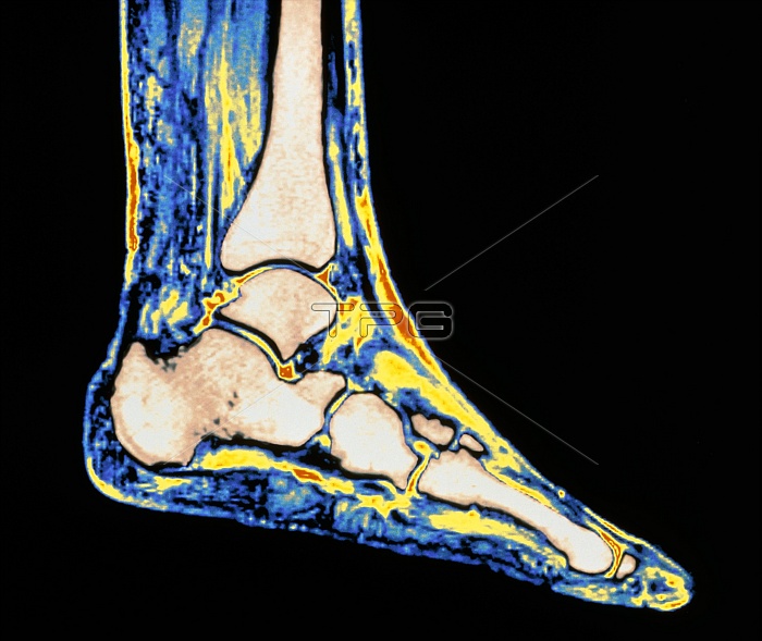
Bones of the ankle. Magnetic Resonance Imaging (MRI) scan of a sagittal (side) view through the healthy ankle of a woman aged 58. The ankle forms a hinge joint between the leg (at top) and foot (lower frame). Bones are beige coloured; soft tissue is brightly coloured. The tibia is the main leg bone seen; its concave head articulates with the cube-shaped talus, the uppermost bone in the foot. The talus is above the calcaneus bone which forms the heel projection behind the foot. The talus and calcaneus attach to tarsal bones in the middle of the foot, which in turn articulate with long metatarsal bones at the base of the toes.
| px | px | dpi | = | cm | x | cm | = | MB |
Details
Creative#:
TOP10217081
Source:
達志影像
Authorization Type:
RM
Release Information:
須由TPG 完整授權
Model Release:
N/A
Property Release:
N/A
Right to Privacy:
No
Same folder images:
 Loading