
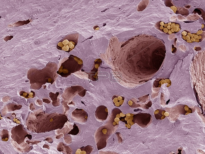
Fossilised compact bone. Coloured scanning electron micrograph (SEM) of a section through fossilised compact bone. This tissue is found in the dense walls of the shafts of bones. It consists of concentric layers of collagen- containing matrix (lamellae, pink), around Haversian canals (large holes), which contain blood and lymph vessels, and nerves. These canals run the length of the bone. The lamellae and canals together form a Haversian system, or osteon. Many systems are arranged in columns running parallel to the long axis of the bone. Mineral deposits (yellow) have collected in the lacunae (smaller holes) of the bone during fossilisation.
| px | px | dpi | = | cm | x | cm | = | MB |
Details
Creative#:
TOP10216970
Source:
達志影像
Authorization Type:
RM
Release Information:
須由TPG 完整授權
Model Release:
N/A
Property Release:
N/A
Right to Privacy:
No
Same folder images:
 Loading