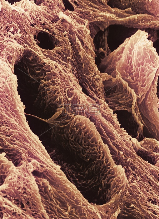
Foetus compact bone. Coloured scanning electron micrograph (SEM) of a section through the develop- ing bone in the foot of a human foetus. This region, known as the epiphysis, is at the end of the long bone within a foetal foot. It will develop separately from the main shaft of the bone, but will eventually fuse to form a complete bone. The dark areas at lower left & upper right are Haversian canals, an interconnecting neurova- scular network containing blood vessels and nerves. The small cavities on the bone's surface are canaliculi. These are interconnecting canals which allow circulation of tissue fluid and the circulation of metabolites. Magnification unknown.
| px | px | dpi | = | cm | x | cm | = | MB |
Details
Creative#:
TOP10216905
Source:
達志影像
Authorization Type:
RM
Release Information:
須由TPG 完整授權
Model Release:
N/A
Property Release:
N/A
Right to Privacy:
No
Same folder images:

 Loading