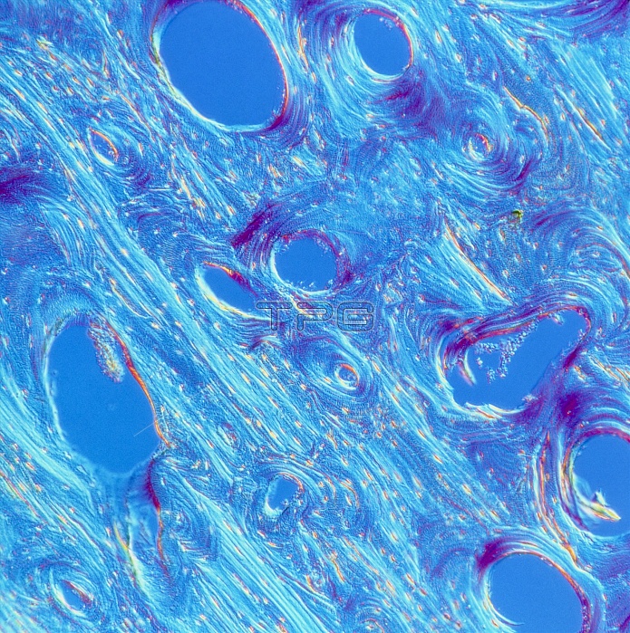
Compact bone. Light micrograph of a section through compact bone from a human femur (thigh bone). This type of bone comprises the dense walls of the shafts of long bones. Compact bone is arranged into concentric bony layers (lamellae) arranged around channels (Haversian canals, seen here as clear areas) containing blood, lymph vessels and nerves. The lamellae and canals form a Haversian system. The Haversian systems are arranged in columns which run parallel to the long axis of the bone. The small lighter-coloured ovals are spaces (lacunae) which house osteocytes, the cells responsible for the maintenance of the bony matrix. Phase contrast. Magnification unknown.
| px | px | dpi | = | cm | x | cm | = | MB |
Details
Creative#:
TOP10216900
Source:
達志影像
Authorization Type:
RM
Release Information:
須由TPG 完整授權
Model Release:
N/A
Property Release:
N/A
Right to Privacy:
No
Same folder images:

 Loading