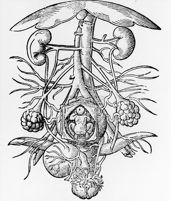
Anatomy of pregnancy. Engraving from the 16th cen- tury of the anatomy of a pregnant woman's urogeni- tal system. Within the opened uterus (lower cent- re) is a foetus. The ovaries are at lower left & lower right, with the vagina at bottom centre. At bottom left is the bladder, whilst the kidneys are at upper left and upper right. The vertical blood vessels between the kidneys are the aorta artery (narrower of the 2) and the inferior vena cava vein. These split (at centre) to form iliac veins & arteries. The umbrella-shaped object (at top) is the liver. Image taken from De conceptu et generatione hominis (1580) by Jakob Rueff.
| px | px | dpi | = | cm | x | cm | = | MB |
Details
Creative#:
TOP10216374
Source:
達志影像
Authorization Type:
RM
Release Information:
須由TPG 完整授權
Model Release:
N/A
Property Release:
N/A
Right to Privacy:
No
Same folder images:

 Loading