
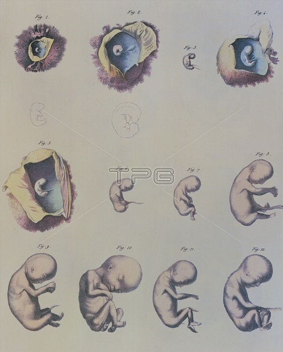
Human embryo. Composite artwork showing 12 images of a developing human embryo. Four images (1, 2, 4 & 5) show the embryo inside the womb. The embryos in Figures 1 and 2 have a larger line drawing beneath them to show more detail. The long struct- ure attached to the belly of the embryos is the umbilical cord which delivers food and oxygen to the embryo from the mother. In the early stages of development, different areas of the embryo become differentiated to form the various struct- ures and organs. In later stages these structures develop, making the embryo more recognisable as a human. Image taken from Manuel D'Anatomie de Corps Humain (1825) by Jules Cloquet. Age of different stages unknown.
| px | px | dpi | = | cm | x | cm | = | MB |
Details
Creative#:
TOP10216373
Source:
達志影像
Authorization Type:
RM
Release Information:
須由TPG 完整授權
Model Release:
N/A
Property Release:
N/A
Right to Privacy:
No
Same folder images:
 Loading