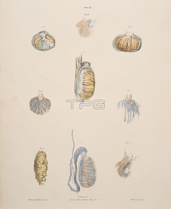
Testis. Historical illustrations of the anatomy of the testis (male sex organ) and its ducts. At upper left, right and centre, the lobed tissue (yellow) of the testis is shown in its capsule. It is seen in isolation at lower left. At centre left, centre right and bottom centre are sections showing the seminiferous tubules (blue) which produce the male sex cells (spermatozoa). These are stored in the epididymis (upper left of testis in bottom centre diagram) and ejaculated through the vas deferens tube (also blue). The descent of the testes (blue) outside the foetal body is shown at top and at lower right. Colour lithograph by Fairland from The Viscera of the Human Body, 1840.
| px | px | dpi | = | cm | x | cm | = | MB |
Details
Creative#:
TOP10216362
Source:
達志影像
Authorization Type:
RM
Release Information:
須由TPG 完整授權
Model Release:
N/A
Property Release:
N/A
Right to Privacy:
No
Same folder images:

 Loading