
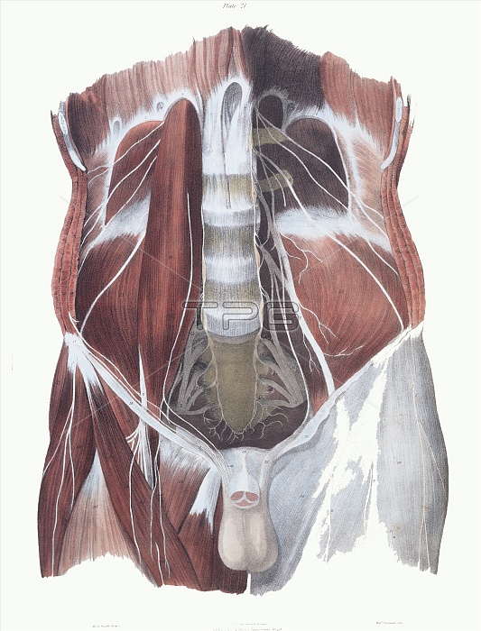
Abdominal spinal nerves. Historical anatomical artwork of the spinal nerves (white) in a human abdomen. This view, from the front, is obtained by removing most of the abdominal organs to reveal the muscles (red) of the back of the abdomen. The lumbar (lower) spine is seen at centre. At left is the right psoas muscle. The left psoas muscle has been removed to show a network of nerves (right) from the spinal cord. This network is called the lumbar plexus. Lower down is another network, the sacral plexus (both sides seen). The largest nerve seen in the lumbar plexus is the crural nerve (the one towards the leg). Artwork from The Nerves of the Human Body (Ed. Jones Quain, London, 1839).
| px | px | dpi | = | cm | x | cm | = | MB |
Details
Creative#:
TOP10216298
Source:
達志影像
Authorization Type:
RM
Release Information:
須由TPG 完整授權
Model Release:
N/A
Property Release:
N/A
Right to Privacy:
No
Same folder images:
 Loading