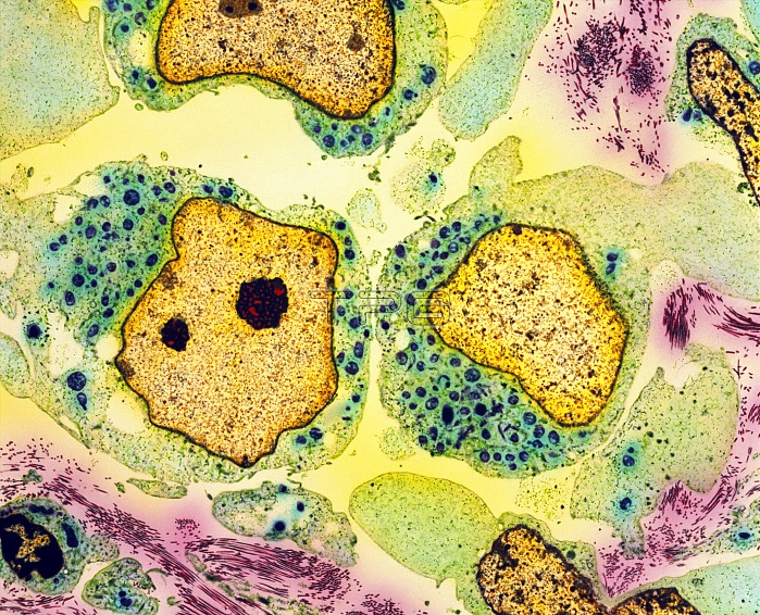
Testicular cancer. Coloured transmission electron micrograph (TEM) of a section through teratoma cancer cells in a testis. Three rapidly-dividing cancer cells are seen at centre left, centre right and lower right. They have large, irregular nuclei (pale brown) and green cytoplasm. Malignant tera- toma of the testis mostly affects young men. They are thought to develop from cells misplaced during embryonic development. Diagnosis is confirmed by removal of the testis and microscopic examination. This may effect a cure, but radiotherapy and anticancer drugs often follow to stop the cancer spreading through the body. 95-100% of cases detected early are cured. Magnification unknown.
| px | px | dpi | = | cm | x | cm | = | MB |
Details
Creative#:
TOP10214804
Source:
達志影像
Authorization Type:
RM
Release Information:
須由TPG 完整授權
Model Release:
N/A
Property Release:
N/A
Right to Privacy:
No
Same folder images:

 Loading